
Research

Finding the Genes that Predispose to Trigeminal Neuralgia
|
|
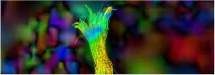
Tumor and White-Matter Modelling
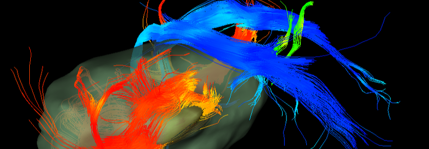
Vestibular schwannoma (acoustic neuroma) are tumors that commonly arise from the vestibular portion of the eighth cranial nerve – the vestibulocochlear nerve. Patients with vestibular schwannoma often present with alterations to the function of this cranial nerve including unilateral sensorineural hearing loss, tinnitus, and imbalance. Stereotactic radiosurgery (SRS) is a successful method of treatment for vestibular schwannoma, helping to reduce their size and restore affected cranial nerve function. However, our understanding of how such cranial nerve function is restored following SRS has been limited due in part to the absence of adequate methods to link microstructural white matter changes along these fiber tracts and restoration of cranial nerve function.
Currently, we are using the technique of diffusion magnetic resonance imaging (dMRI) and tractography to pinpoint microstructural changes in the vestibulocochlear nerve, and other potentially-affected cranial nerves, in patients with acoustic neuromas. We are also linking any such changes with hearing and balance problems or other sensory findings such as facial numbness in these patients. We believe that combining neuroimaging and clinical assessment will ultimately help advance the development of diagnostic methods and effectiveness of SRS. Altogether, we hope to improve clinical outcomes for those with acoustic neuroma-associated cranial nerve dysfunction.
Project Members: Aisha Halawani, Matthew R. Walker, Brendan Behan, Erika Wharton-Shukster, David Q. Chen, Mojgan Hodiae
Funding: Elekta, Mitacs, UHN
Related Publications:
Chen DQ, Quan J, Guha A, Tymianksi M, Mikulis D, Hodaie M. (2011). Three-dimensional in vivo modelling of vestibular schwannomas and surrounding cranial nerves with diffusion imaging tractography. Neurosurg, 68(4): 1077-83.
Hodaie M, Quan J, Chen DQ. (2010). In vivo visualization of cranial nerve pathways in humans using diffusion-based tractography. Neurosurg, 66(4): 788-95.
Balasubramaniam A, Shannon P, Hodaie M, Laperrier N, Michaels H, Guha A. (2007). Glioblastoma multiforme after stereotactic radiotherapy for acoustic neuroma: case report and review of the literature. Neuro Oncol, 9: 447–53.
Structural Abnormalities in Trigeminal Neuralgia
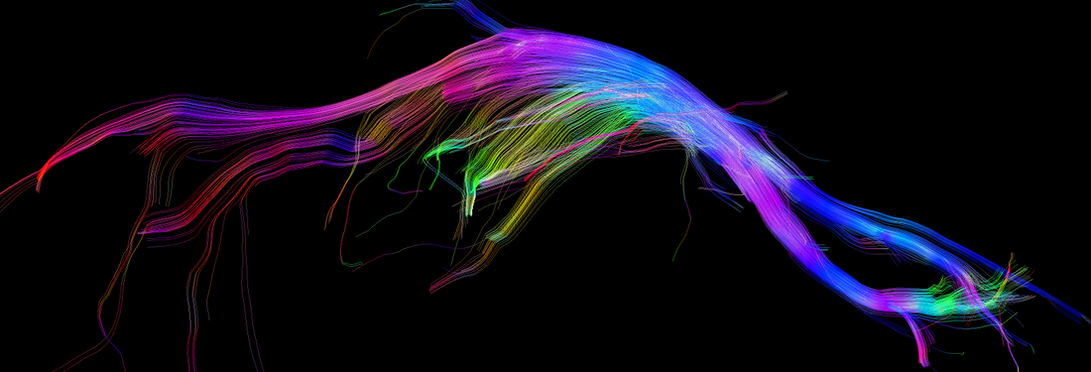
We are interested in using MRI to noninvasively study structural nerve and brain abnormalities in patients with trigeminal neuralgia (TN), a severe neuropathic pain disorder associated with compression of the trigeminal nerve. Specifically, we use diffusion magnetic resonance imaging (dMRI) to analyze trigeminal nerve microstructure, using measures that have been linked to pathophysiological mechanisms such as demyelination and edema. It is now well established that neuropathic pains are also associated with abnormalities in the brain regions involved in the multi-dimensional experience of pain, pain modulation, and motor function, even though they are disorders of a peripheral nerve. Therefore, we measure cortical and subcortical gray matter in the brains of TN patients using cortical thickness analysis and voxel-based morphometry methods, and examine brain white matter microstructure and connectivity using dMRI techniques.
Project members: Peter Shih-Ping Hung, Jidan Zhong, David Q. Chen, Karen Davis (collaborator), Mojgan Hodaie
Funding: PSI Foundation, CIHR
Related publications:
Zhong J, Chen DQ, Hung PS-P, Hayes DJ, Liang KE, Davis KD, Hodaie M. (2018). Multivariate pattern classification of brain white matter connectivity predicts classic trigeminal neuralgia. Pain, 159(10): 2086–2087.
DeSouza DD, Hodaie M, Davis KD. (2016). Structural Magnetic Resonance Imaging Can Identify Trigeminal System Abnormalities in Classical Trigeminal Neuralgia. Front Neuroanat, 10: 95.
DeSouza DD, Hodaie M, Davis KD. (2014). Abnormal trigeminal nerve microstructure and brain white matter in idiopathic trigeminal neuralgia. Pain, 155(1): 37-44.
DeSouza DD, Moayedi M, Chen DQ, Davis KD, Hodaie M. Sensorimotor and Pain Modulation Abnormalities in Trigeminal Neuralgia: a Paroxysmal, Sensory-Triggered Neuropathic Pain. PLoS ONE, 8(6): e66340, 2013.
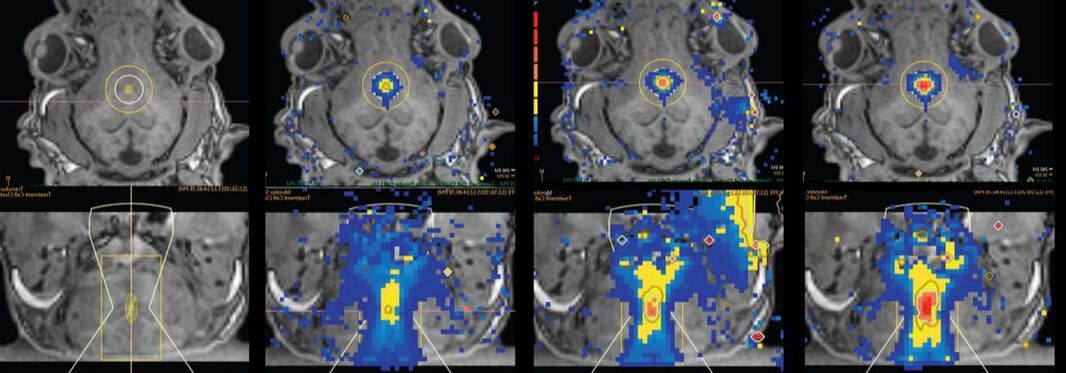
Neurosurgical Treatment-Related Plasticity in Trigeminal Neuralgia Patients
How do neurosurgical interventions for TN affect the nerve and brain? Two common neurosurgical options for TN are microvascular decompression surgery and Gamma Knife radiosurgery. Although these interventions target different regions of the trigeminal nerve and presumably have different mechanisms of action, they are both associated with high degree of pain relief. Our goals are to determine the neural correlates of this pain relief to gain insight into how these interventions produce analgesia and also to develop potential biomarkers for effective treatment.
Project members: Peter Shih-Ping Hung, David Q. Chen, Karen Davis (collaborator), Mojgan Hodaie
Funding: PSI Foundation, CIHR
Related publications:
Hung PS-P, Tohyama S, Zhang JY, Hodaie M. (2019). Temporal disconnection between pain relief and trigeminal nerve microstructural changes after Gamma Knife radiosurgery for trigeminal neuralgia. J Neurosurg, Epub 2019 Jul 12:1–9.
Tohyama S, Hung PS-P, Zhong J, Hodaie M. (2018). Early postsurgical diffusivity metrics for prognostication of long-term pain relief after Gamma Knife radiosurgery for trigeminal neuralgia. J Neurosurg, 131(2): 539-548.
Al-Otaibi F, Alhindi H, Alhebshi A, Albloushi M, Baeesa S, Hodaie M. (2013). Histopathological effects of radiosurgery on a human trigeminal nerve. Surg Neurol Int, 4(6): S462-S467.
Hodaie M, Coello AF. (2013). Advances in the management of trigeminal neuralgia. J Neurosurg Sci, 57(1): 13-21.
Hodaie M, Chen DQ, Quan J, Laperriere N. (2012). Tractography delineates microstructural changes in the trigeminal nerve after focal radiosurgery for trigeminal neuralgia. PLoS One, 7(3): e32745.
Neuroimaging and Emotion in Chronic Neuropathic Facial Pain
Chronic neuropathic pain is caused by injury to the nervous system and is estimated to affect between 2-8% of the total population. It has a tremendous impact on Canadian society with its negative effects on health, quality of life, increased suicidality and loss of work-place productivity and associated costs estimated at $40 billion. Trigeminal neuralgia and postherpetic neuralgia are two examples of chronic neuropathic pain syndromes which result in extreme facial pain. Brain imaging studies in people with chronic pain have shown differences in the architecture of the brain associated with physical touch and emotion. However, no studies have looked at the connection between emotion-related brain circuits and behaviour in syndromes of facial neuropathic pain. By combining different brain imaging techniques and psychological tests, the goal of this study is to identify the relationship between the architecture of emotional brain circuits and related behaviour in people with these syndromes. We believe that differences in behaviour will predict brain-based differences in many emotional brain circuits. This study will uncover the unique brain signatures associated with each of these disorders and may help us predict who will respond to treatment, which is key for developing a personalized medical approach.
Project members: Peter Shih-Ping Hung, Erika Wharton-Shukster, Dave J. Hayes, David Q. Chen, Karen Davis (collaborator), Mojgan Hodaie
Related publications:
Hayes DJ, Chen DQ, Zhong J, Lin A, Behan B, Walker M, Hodaie M. (2017). Affective circuitry alterations in patients with trigeminal neuralgia. Front Neuroanat, 11:73.
DeSouza DD, Davis KD, Hodaie M. (2015). Reversal of insular and microstructural nerve abnormalities following effective surgical treatment for trigeminal neuralgia. Pain, 156(6):1112-23.
Rosen A, Chen DQ, Hayes DJ, Davis KD, Hodaie M. (2015). A neuroimaging strategy for the three-dimensional in vivo anatomical visualization and characterization of insular gyri. Stereotact Funct Neurosurg, 93(4):255-64.
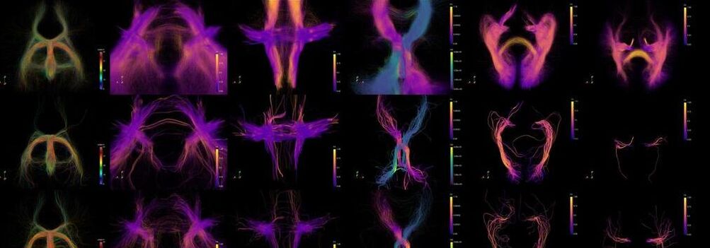
Cranial Nerve Tractography and Tractographic Analysis Development
Project members: David Q. Chen, Karen Davis (collaborator), Mojgan Hodaie
Funding: PSI Foundation, CIHR
Related publications:
Behan B, Chen DQ, Sammartino F, DeSouza DD, Wharton-Shukster E, Hodaie M. (2017). Comparison of diffusion-weighted MRI reconstruction methods for visualization of cranial nerves in posterior fossa surgery. Front Neurosci, 11:554.
Chen DQ, Zhong J, Hayes DJ, Behan B, Walker M, Hung PS, Hodaie M. (2016). Merged group tractography evaluation with selective automated group integrated tractography. Front Neuroanat, 10:96.
Hodaie M, Chen DQ, Quan J, Laperriere N. (2012). Tractography delineates microstructural changes in the trigeminal nerve after focal radiosurgery for trigeminal neuralgia. PLoS One, 7(3): e32745.
Hodaie M, Quan J, Chen DQ. (2010). In vivo visualization of cranial nerve pathways in humans using diffusion-based tractography. Neurosurg, 66(4): 788-95.
Neuroimaging Correlates of Pain in Multiple Sclerosis Patients with Trigeminal Neuralgia
Project members: David Q. Chen, Paul O'Connor (collaborator), Karen Davis (collaborator), Mojgan Hodaie
Funding: Multiple Sclerosis of Canada
Related publications:
Chen DQ, DeSouza DD, Hayes DJ, Davis KD, O’Connor P, Hodaie M. (2016). Diffusivity signatures characterize trigeminal neuralgia associated with multiple sclerosis. Mult Scler, 22(1):51-63.
Torres CV, Moro E, Dostrovsky JO, Hutchison WD, Poon Y-YW, Hodaie M. (2010). Unilateral pallidal deep brain stimulation in a patient with cervical dystonia and tremor. J Neurosurg, 113(6): 1230-1233.
Finding the Genes that Predispose to Trigeminal Neuralgia
Trigeminal neuralgia is a sudden, severe, stabbing, and recurring facial pain in one or more branches of the trigeminal nerve. Medications and surgical procedures are available for this condition, but these treatments can have risks and unwanted side effects. The resulting pain relief may be temporary. We need to understand what causes this pain better!
The Hodaie Lab is the sole Canadian site collaborating with Dr. Kim Burchiel at the Oregon Health and Science University (OHSU) to determine whether trigeminal neuralgia has a genetic component. Dr. Burchiel is the Chairman of the Department of Neurological Surgery at OHSU and specializes in functional and stereotactic neurosurgery, pain surgery, and epilepsy surgery. His research focuses on the physiology of pain sensation, neuropathic pains, movement disorders and image-guided neurosurgery.
This is an intensive, multicenter genetic study supported by the Facial Pain Research Foundation. Together, we are striving to find the gene or genes that cause trigeminal neuralgia. Determining this genetic component could potentially improve the diagnosis and treatment of trigeminal neuralgia in the future.
Project members: Erika Wharton-Shukster, Kim Burchiel (collaborator), Mojgan Hodaie
Funding: Facial Pain Research Foundation
Related publications:
Project started in 2014.
Developing a Non-Invasive Treatment of Neurological Disorders using MR-Guided Focused Ultrasound
In collaboration with the Hospital for Sick Children, our goal is to use DWI to identify neurosurgical targets related to paediatric epilepsy and to make technological advancements for the acquisition and analysis of DWI data. Higher order tractography algorithms can be used to visualize relevant pathways at the individual level within both the central and peripheral nervous systems. Quantitative diffusion imaging metrics may also be used for the in vivo assessment of nerve-related changes from treatment and correlations with clinical outcome.
Project members: Matthew R. Walker, Jidan Zhong, David Q. Chen, Dave J. Hayes, James Drake (collaborator), Mojgan Hodaie
Funding: Brain Canada, Mitacs, W. Garfield Weston Foundation
Related publications:
Walker MR, Zhong J, Waspe AC, Looi T, Piorkowska K, Hawkins C, Drake JM, Hodaie M. (2019). Acute MR-guided high-intensity focused ultrasound lesion assessment using diffusion weighted imaging and histological analysis. Front Neurol, 10: 1069.
Boutet A, Ranjan M, Zhong J, Germann J, Xu D, Schwartz ML, Lipsman N, Hynynen K, Devenyi GA, Chakravarty M, Hlasny E, Llinas M, Lozano CS, Elias GJB, Chan J, Coblentz A, Fasano A, Kucharczyk W, Hodaie M, Lozano AM. (2018). Focused ultrasound thalamotomy location determines clinical benefits in patients with essential tremor. Brain, 141(12): 3405-3414.
Zhong J, Chen DQ, Walker M, Waspe A, Looi T, Piorkowska K, Drake JM, Hodaie M. (2016). An in vivo multi-modal structural template for neonatal piglets using high angular resolution and population-based whole-brain tractography. Front Neuroanat, 10:92.
Sammartino F, Krishna V, King NKK, Lozano AM, Schwartz ML, Huang Y, Hodaie M. (2016). Tractography-based ventral intermediate nucleus targeting: Novel methodology and intraoperative validation. Mov Disord, 31(8):1217-25.